Echocardiology Lab
Echocardiography, or echo, is a painless diagnostic test that uses sound waves to create moving pictures of your heart. Echo is performed to
- Show the size and shape of your heart
- Determine how well your heart’s chambers and valves are working
- Show how well blood flows through your heart’s chambers and valves
- Pinpoint areas of heart muscle that aren’t contracting well because of poor blood flow or injury from a previous heart attack
- Detect possible blood clots inside the heart, fluid buildup in the pericardium (the sac around the heart), and problems with the aorta
Echo Tests Performed at Crouse Health
Stress Test
A stress test, often called a treadmill test, measures how your heart works when experiencing added workload or “stress” of exercise.
Stress Echocardiogram
A stress echo is a more dynamic test that examines the heart in action. It combines an ultrasound of the heart with a stress test.
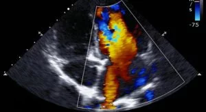
2-D Echocariogram
This two-dimensional technique is used to “see” the actual motion of the heart structures. A 2-D echo view appears cone-shaped on the monitor, and the real-time motion of the heart’s structures can be observed. This enables the doctor to see the various heart structures at work and evaluate them.
Transesophageal Echocardiogram
A transesophageal echo (TEE) test is a type of echo that uses a long, thin, tube (endoscope) to guide the ultrasound transducer down the esophagus, or the “food pipe” that goes from the mouth to the stomach. A TEE provides pictures of the heart without the ribs or lungs getting in the way.