Contrast-Enhanced Mammography
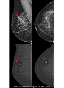
Top: Views of a standard digital mammogram with cancer (red arrows), which is difficult to see, even for a trained radiologist. Bottom: “iodine-only” CEM images in the same patient, where the mass due to invasive cancer (arrows) is clearly visible.
What is Contrast-Enhanced Mammography?
Contrast-Enhanced Mammography (CEM) is a standard, digital mammogram. The main difference is CEM uses an injection of contrast (similar to CT) prior to imaging. This injection will go through an IV in your arm or hand and will highlight abnormal findings in your breast tissue that may not be able to be seen on a standard mammogram. Positioning and compression of the breast are the same as a standard mammogram.
Why am I having a CEM?
CEM can be used to assess new or unexplained breast symptoms or as an additional workup for abnormalities seen on standard mammograms.
What are the benefits of CEM?
Contrast-Enhanced Mammography can help find areas in your breast where cancer may not be seen on a conventional mammogram. It’s a vascular study, like an MRI, but can be used in cases where MRI cannot be tolerated. It is cost-effective and produces higher-resolution images.
What are the risks of CEM?
There may be a possible allergic reaction to the contrast dye used during the exam.
How to Prepare for CEM
Bloodwork may be required before your exam if you:
- are over 60 years old
- have diabetes or renal disease
- take Metformin or any metformin-containing medications
Before your exam you should make sure to hydrate. Do not wear any deodorants, powders or lotions. You will be able to drive yourself to and from the appointment. If you choose to bring someone with you, they must wait in the waiting room.
After CEM
One of our breast radiologists will review your exam and any abnormalities seen may be recommended for ultrasound, biopsy or MRI.